3Rs in action: Can organoids replace stressful fasting models in animal research?
For some animal experiments, animals must fast. A pilot project funded by the UZH Office for Animal Welfare and 3R is investigating whether intestinal organoids can replace these burdensome animal experiments.
At the Department of Molecular Life Sciences of the University of Zurich, a pilot project funded through the AW&3R Grant by the Office for Animal Welfare and 3R explored whether intestinal organoids could replace animal fasting experiments used to study how cells respond to nutrient availability. Intestinal organoids are tiny 3D structures grown in the lab from stem cells that behave comparable to the intestine. These are used to study digestion, diseases, and medicines without involving living animals.
Understanding how cells regulate protein production is essential for research into tissue function, metabolism, and disease. Previous studies showed that in the mouse intestine, messenger RNAs, small molecules that carry instructions from DNA to the cell, are positioned within cells in ways that may help fine-tune protein synthesis during fasting. However, these experiments rely on fasting mice during their natural active feeding period, a physiologically stressful intervention.
The goal of this project, led by Prof Franka Voigt was to develop a laboratory-grown alternative that could reproduce these cellular responses without the use of fasted animals.
From animal models to organoids
Organoids are miniature tissue models grown from stem cells that replicate key features of real organs. In this project, researchers focused on intestinal organoids that mimic the lining of the gut.
A major challenge was that standard commercial culture media did not generate organoids mature enough to reflect the organization of native intestinal tissue. Cell differentiation remained incomplete, making it difficult to study how specific cell types regulate protein production.
To overcome this, the team developed an optimized culture medium with a defined combination of growth factors. Growth factors are natural substances that tell cells to grow, divide, or change. They help control how cells develop and heal.
How the system works
Under the improved conditions, organoids developed a cell-type composition more closely resembling the natural intestinal epithelium.
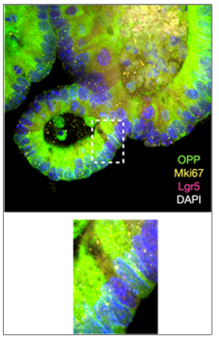
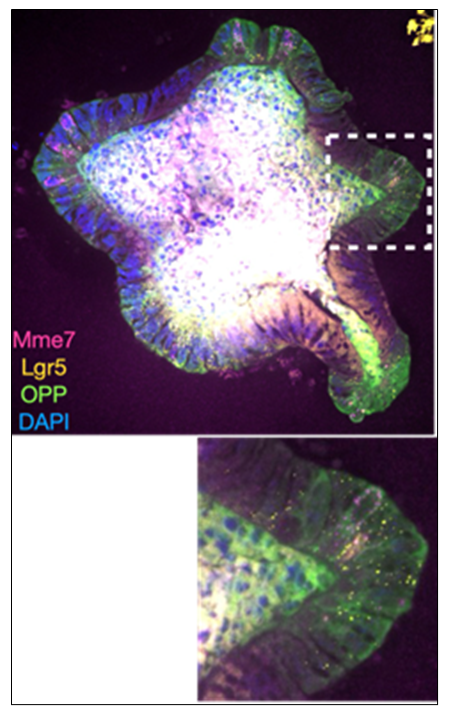
Then the researchers used a method called “clickable puromycin” (OPP labeling) to make newly made proteins visible and measure how active protein production is – the process by which cells build proteins from RNA. The signal was very specific: it appeared when puromycin was added, and became much weaker when ribosomes (the cell’s protein-making machinery) were blocked with drugs.
It is also important that the patterns of protein production matched what has been seen in animal studies. High activity was found in areas similar to intestinal folds where many stem cells are located, while more mature cell areas showed lower activity. These results suggest that optimized organoids can reproduce important biological differences between cell states that were previously studied in animals.
Advancing the 3Rs through innovation
This project contributes primarily to the Replacement and Reduction principle of the 3Rs (Replace, Reduce, Refine):
Replacement: Organoids offer a promising non-animal model for studying fasting-related cellular responses if based on human cells. They replace burdening animal experiments. In this project mouse cells were used to culture organoids.
Reduction: More predictive in vitro systems may reduce the need for exploratory animal studies in future projects.
A promising foundation
Further validation under nutrient-rich and nutrient-poor conditions is still required, and the findings remain preliminary. Nevertheless, the project establishes an important foundation for replacing stressful in vivo fasting models with robust organoid-based alternatives.